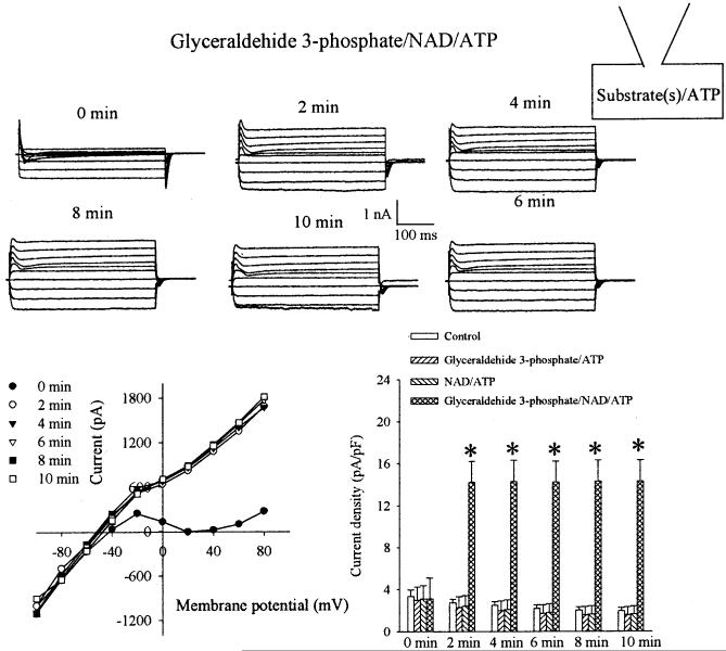
FIG. 6.
Substrates of GAPDH regulate the activity of sarcolemmal KATP channels. Membrane currents were recorded using conventional patch-clamp electrophysiology and corresponding I-V relationships (each point is the means ± SE, n = 5) at depicted conditions (each substrate was added to the pipette solution at a concentration of 20 mmol/l, whereas ATP was kept at 5 mmol/l). Time point 0 min refers to the recording made immediately after the whole-cell configuration was established. The bar graph shows current density at +80 mV in cells under depicted conditions and time points. Each bar represents the means ± SE (n = 4-11). *P < 0.01 when compared with the control.

