
Figure 6.
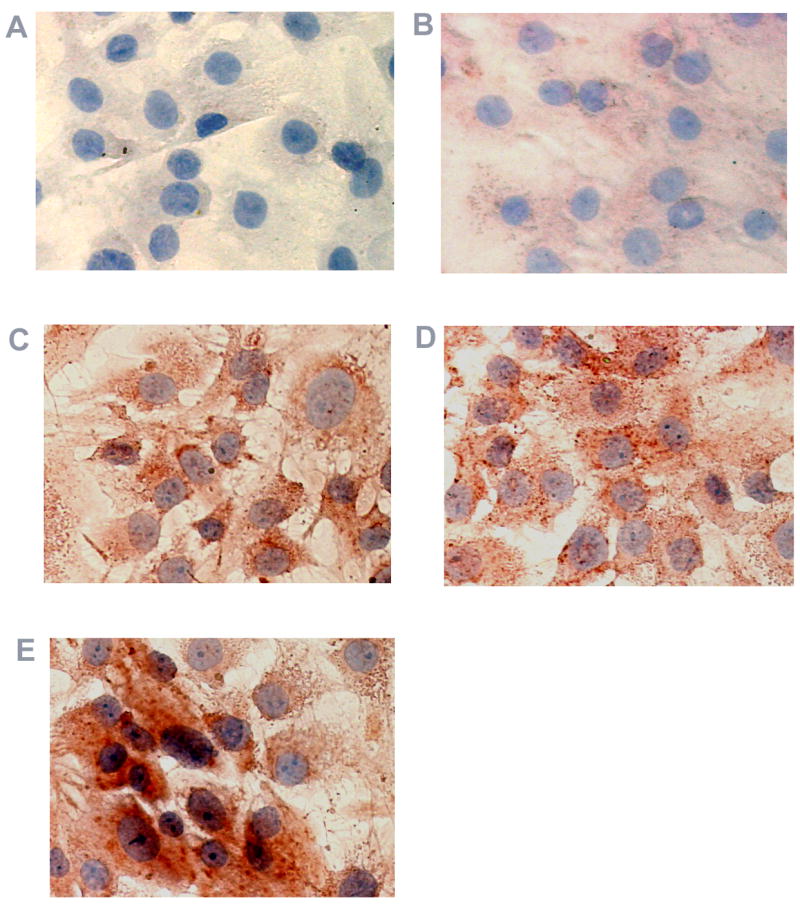
Immunohistochemical analysis of VEGF protein expression in cultured human RPE cells. RPE cells were exposed to thrombin, TGF- β2, or thrombin plus TGF- β2 for 16 hr. Cells in (A) were stained with normal rabbit serum serving as non-specific control. Untreated RPE cells (B) and cells treated with thrombin (C), TGF- β2 (D), and thrombin plus TGF- β2 (E) were stained using rabbit anti-human VEGF antibody and the red chromogen, AEC.