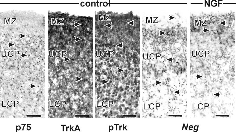
Figure 6. Expression of neurotrophin receptors and neg at P3.
Immunohistochemical labeling of the neurotrophin receptors showed that p75-positive cells were only localized in the cortical plate, not in the marginal zone. In contrast, trkA- and phosphorylated trk-positive cells were apparent throughout the cortical wall. In situ hybridization for Neg labeled cells in all three segments of the cortical wall. Treatment with NGF did not alter Neg expression. Nomenclature as for Figure 5. Scale bars are 50 μm.

