
Figure 3.
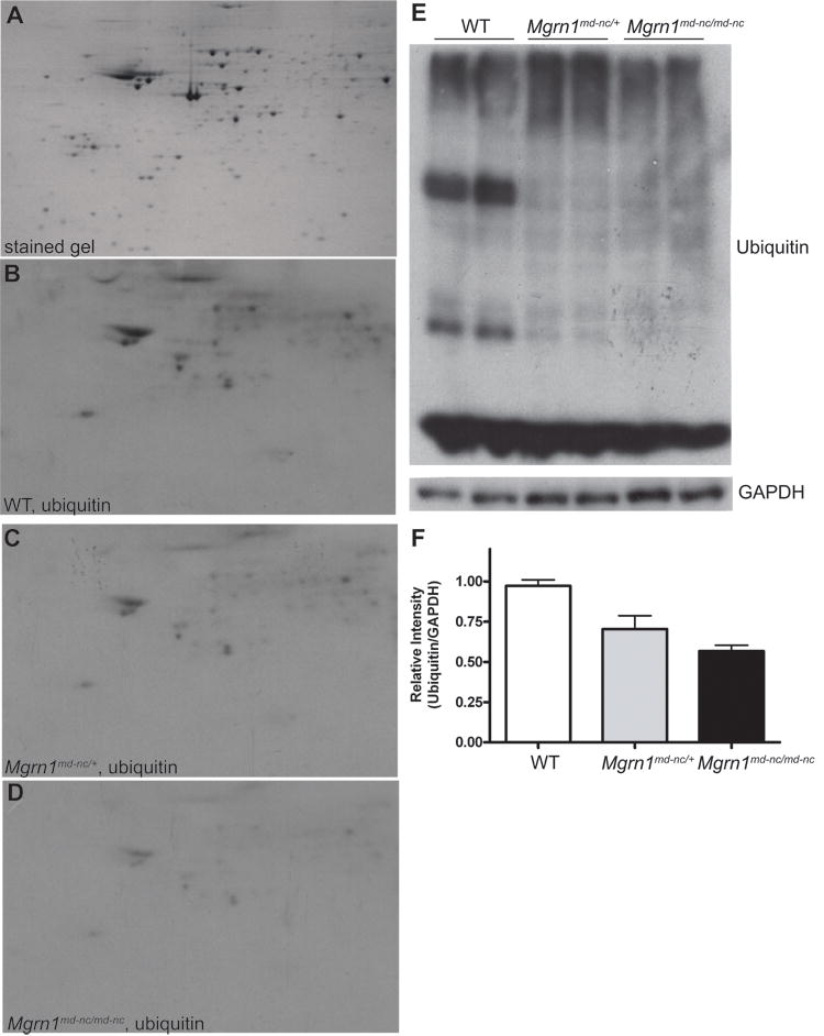
Dose-dependent, global reduction in ubiquitination associated with MGRN1-deficiency. (A) Representative 2-DG gel of complete brain proteome stained with colloidal Coomassie blue. (B-D) Western analysis of ubiquitin complete brain protein extracts from 6-month old wild-type (WT) (B), Mgrn1md-nc/+ (C) and Mgrn1md-n/md-nc (D) mice following 2-DGE. (E) 1-DGE analysis of ubiquitin levels in brain protein extracts from 6-month old wild-type (WT), Mgrn1md-nc/+ and Mgrn1md-n/md-nc mice. Blots were immunoblotted for ubiquitin, then stripped and immunoblotted for GAPDH as a control for protein loading. Signal intensity was quantified using ImageJ software and plotted as average ratio of ubiquitin signal/GAPDH signal by genotype (F) to quantify differences in protein ubiqutination. Ratios were normalized against WT.