
Figure 6.
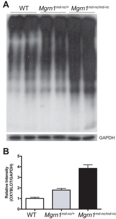
Elevated oxidative damage to brain proteins in Mgrn1 null mutants. (A) Oxyblot (top panel) assay of brain lysates from 6-month old mice revealed increased levels of protein carbonyls in Mgrn1md-nc/md-nc mutants relative to Mgrn1md-nc/+ siblings. Blots were stripped and blotted for GAPDH as loading control (bottom panel). Signal intensity was quantified using ImageJ software and plotted as average ratio of the Oxyblot signal/GAPDH signal by genotype (B) to quantify differences in oxidatively damaged proteins. Ratios were normalized against WT.