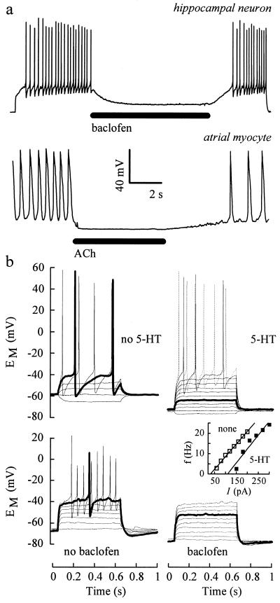
Figure 4.
GIRK activation inhibits action potential firing. (a) Bars indicate the application of 50 μM baclofen to a 13 d-cultured neuron (3 d after coinfection with AdGIRK1+2), and of 5 μM ACh to an atrial cell. A 15.8 sec current pulse of +60 pA was injected into the neuron to cause firing; the myocyte was firing spontaneously. Resting EM were −70 mV (neuron) and −66 mV (myocyte). (b) EM responses of AdGIRK1+2-coinfected neurons to 0.6 sec current pulses in the presence (Right) and absence (Left) of 30 μM 5-HT (Upper) and 50 μM baclofen (Lower); steps of 20 pA (Upper, from −20 pA) and 16 pA (Lower, from 0 pA). Note that the 60 and 96 pA pulses cause action potential firing in the absence but not in the presence of 5-HT and baclofen, respectively (thick lines). (Inset) Firing frequencies calculated from action potential intervals of the top cell in the presence (▪) and absence (□) of 5-HT. Solid lines show linear fits; x axis intercepts indicate ITh.

