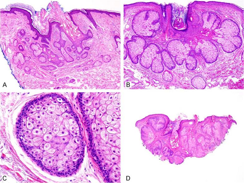
Figure 1.

Sebaceous hyperplasia shows superficial sebaceous lobules surrounding a central pore (A, B). Higher power reveals foamy eosinophilic cytoplasm in central cells and only two basaloid or germinative cell layers at the periphery (C). (D) While most of the lesion appears to be sebaceous hyperplasia, a few of the lobules have expanded basaloid areas, indicating that this lesion is best considered as sebaceous adenoma.
