Figure 2.

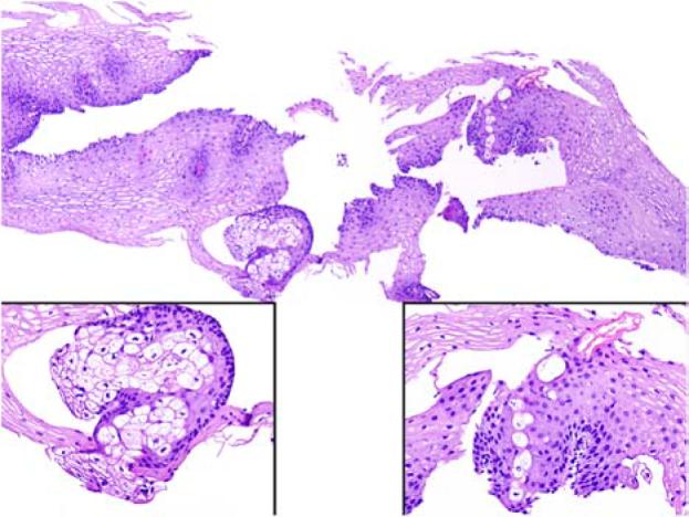
Ectopic sebaceous gland in an oesophageal mucosa biopsy. The left and right insets magnify the sebaceous lobule and sebaceous ductal structure (courtesy of Dr. Huamin Wang, UT MD Anderson Cancer Center, Houston, Texas, USA).

Ectopic sebaceous gland in an oesophageal mucosa biopsy. The left and right insets magnify the sebaceous lobule and sebaceous ductal structure (courtesy of Dr. Huamin Wang, UT MD Anderson Cancer Center, Houston, Texas, USA).