
Figure 2.
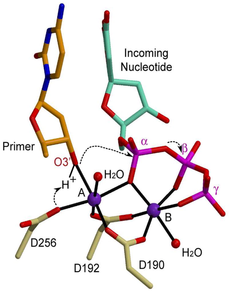
Catalytic mechanism for nucleotidyl transfer. The primer terminal residue (orange) is drawn in stick, with the O3′ in red. The incoming nucleotide is cyan, with its phosphates in magenta. Protein residues from the ternary structure of Pol β (PDB code 2FMS) are khaki. Magnesium ions are purple and water molecules completing coordination of the metal ions are small red spheres.