
Fig 5.
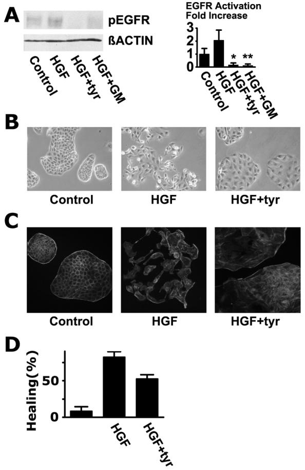
Transactivation of the EGFR is required for induction of a fully motile phenotype in MDCK cells. (A) Activation of the EGFR in MDCK cells under conditions similar to those shown in Fig. 1B except that 20 ng/ml HGF was used. The signals were significantly reduced in the presence of tyrphostin AG1478 and GM6001 (*, P< 0.0005; **, P< 0.001) (B) Phase contrast photo-micrographs of control MDCK colonies, colonies stimulated with 20 ng/ml HGF for 7 hours, and colonies stimulated with HGF in the presence of 10 μM tyrphostin AG 1478 (tyr). (C) Cells were stained with Alexa Fluor® 546-labeled phalloidin. (D) Wounding assay was performed as in Fig. 3B. The means of quadruplicate measurements are shown and the error bars are standard deviations.