
Fig. 5.
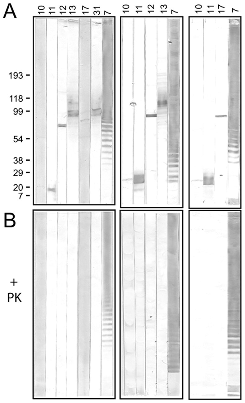
Proteinase K (PK) sensitivity of target antigens of anti-LVS hybridoma antibodies. One half of a sample of LVS lysate was treated with PK, then each the untreated and PK-treated samples were subjected to SDS-PAGE under reducing conditions and transferred to nitrocellulose membranes. The membranes were cut into strips and each strip probed with the indicated hybridoma antibody. Ab7, an LPS-binder, was used as negative control for PK-treatment. The untreated (A) and PK-treated (B) membrane strips for each antibody were developed simultaneously and for the same time. Immunoblots from three separate experiments are shown; the nitrocellulose membranes in the middle and right immunoblots were “aged” for two weeks prior to use to allow visualization of the bands identified by antibodies 10 and 17. The positions of molecular weight markers (in kDa) are indicated for the left immunoblot.