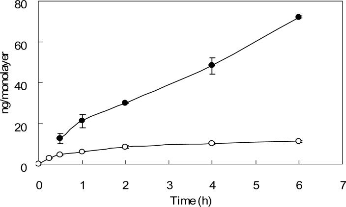
Fig. 3. Cellular uptake of 125I-Tf in confluent and subconfluent Caco-2 cell monolayers.
Caco-2 cell monolayers with approximately 25% confluence were used as subconfluent cells. Confluent (solid circles) and subconfluent (open circles) Caco-2 cell monolayers on 12-well cluster plates were pre-incubated with serum free medium at 37°C for 1 h to deplete serum Tf. Subsequently, 125I-Tf was added to cells and incubated for different time intervals at 37°C. Cells were then washed with cold PBS and solubilized by incubation with 1 N NaOH. The cell lysate was assayed by using a Packard gamma counter. Non-specific binding was determined in parallel. Each point represents the mean of three measurements with error bars representing the standard deviation.

