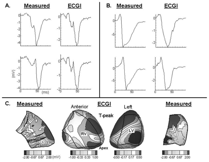
Figure 2.

Panel A, invasively measured and noninvasively reconstructed electrograms from the mid to basal region and the right margin of the RV. Panel B, invasively and noninvasively reconstructed electrograms from remote sites closer to the interventricular septum. Panel C, invasively measured (first and last column) and noninvasively reconstructed epicardial potentials during repolarization. Boundaries of the intraoperative 200 electrode sock are overlaid over the RV and LV.
