FIG. 2.

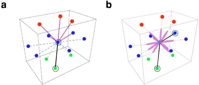
(Color online) Lattice imposed constraints for the nucleic acid chain configurations. (a) We use a three-dimensional triangular lattice. We constrain consecutive phosphodiester bonds to have an angle larger than or equal to 60° in order to mimic the stiffness of the sugar-phosphate backbone. The diagram illustrates the conformations allowed for two consecutive bonds in a three-dimensional triangular lattice. The black solid line and the black circles represent the reference bond and beads, respectively, and the purple dashed lines indicate the allowed conformations for the following bond. In the diagram, colored dots represent lattice sites which are nearest neighbors of the central blue dot. Different colors indicate the plane on which the site sits: top (red), middle (blue), and bottom (green). (b) The phosphodiester bond can be easily torqued, hence a sugar-base complex can take any spatial orientation provided it does not overlap with the phosphodiester bond. The diagram illustrates the ten possible orientations that a base (pin) can take (purple ellipses) for a given conformation of the polymer chain indicated by the black circles and black solid lines.
