
Figure 1. srp-6 Nulls Displayed the Osl Phenotype.
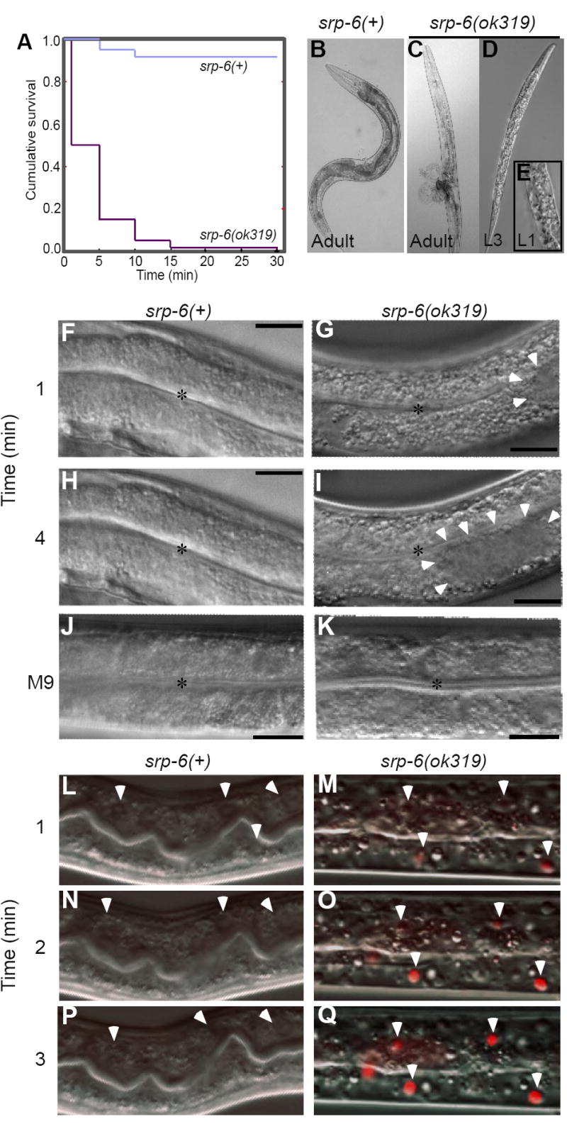
(A) Kaplan-Meier survival curves of srp-6(+) and srp-6(ok319) animals after immersion in 25 °C water (n ≈ 300 animals/group; P < 0.001, log-rank test).
(B-K) Morphology of srp-6(+) (B, F, H, J) and srp-6(ok319) (C-E, G, I, K) animals during hypotonic stress. Low resolution DIC images of adults (B, C) and larvae (D, E) after 5 min in water. The L1 was fixed in 3% formaldehyde. For higher resolution DIC imaging (F-K; bar = 50 μm), L4s were immersed in water (F-I) or M9 (J, K). Intestinal cell cytoplasm of srp-6(ok319) animals was refractile (G, I) and clear cytoplasmic vacuoles (arrowheads) expanded and coalesced (*intestinal lumen).
(L-Q) Intestinal cell plasma membrane integrity was lost in srp-6(ok319) (M, O, Q), but not in srp-6(+) (L, N, P) animals after exposure to water. Confocal microscopy was used to monitor propidium iodide uptake over time. Arrowheads indicate intestinal cell nuclei in dying animals.