
Figure 5. Instability of Lysosomal Gut Granules in Osl.
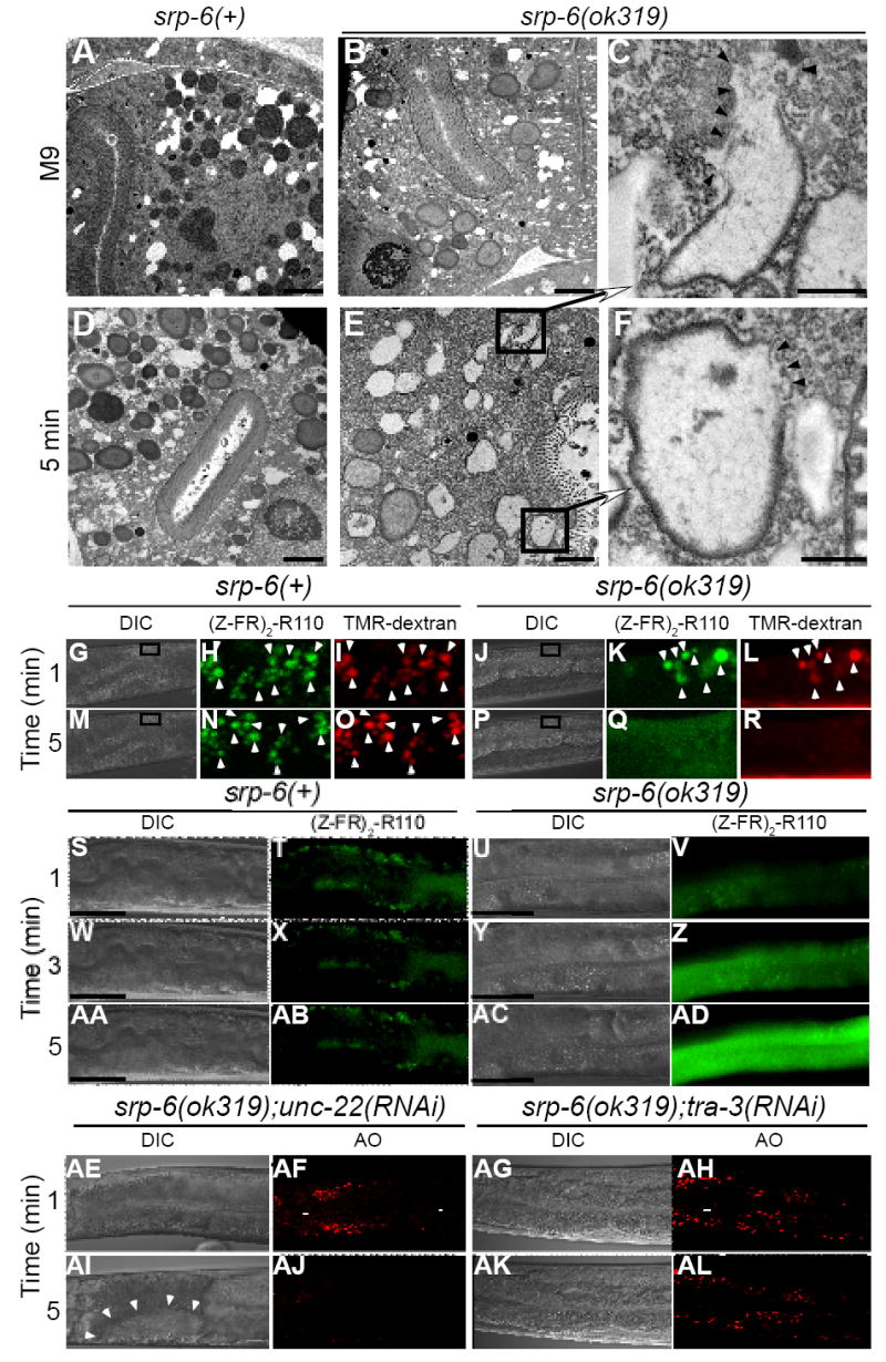
(A-F) Transmission electron microscopy of srp-6(+) (A, D) or srp-6(ok319) (B, E) animals 5 min after immersion in M9 (A, B) or water (D, E) (bar = 2 μm). Higher magnification (C, F) of lysosomal-like granules in (E) (bar = 500 nm). Arrowheads demarcate loss of granule membranes.
(G-R) Live cell confocal microscopic imaging of the fluorescent cysteine peptidase substrate, (Z-FR)2-R110 (H, K, N, Q) and the fluorescent endocytic maker, TMR-dextran (I, L, O, R) in srp-6(+) (G-I, M-O) or srp-6(ok319) (J-L, P-R) animals after immersion in water. The fluorescent images were magnified from a DIC image (boxed inset) within the intestinal cell cytoplasm (G, M, J, P). The lysosomal-like gut granules of both srp-6(ok319) and srp-6(+) animals acquired both labels (arrowheads) and were mostly coincident in the merged images (Movies 5A and 5B).
(S-AD) Live widefield DIC and fluorescence microscopy of srp-6(+) and srp-6(ok319) animals labelled with (Z-FR)2-R110 and immersed in water. Fluorescent gut lysosomes in the srp-6(+) animal remained intact (T, X, AB; bar = 50 μm). In the srp-6(ok319) animals, the disappearance of fluorescent lysosomes was accompanied by a transient wave of intense cytoplasmic fluorescence that propagated down the intestinal cell (V, Z, AD; Movies 6A and 6B).
(AE-AL) Live DIC (AE, AG, AI, AK) and confocal microscopy (AF, AH, AJ, AL) of AO-labelled srp-6(ok319);unc-22(RNAi) (AE, AF, AI, AJ) or srp-6(ok319);tra-3(RNAi) (AG, AH, AK, AL) animals immersed in water. Vacuoles are indicated by arrowheads.