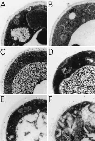
Figure 6.
Immunoelectron localization of Pir proteins to the cell wall. Thin sections of cells of strains BWG7a (A and B), DT8–1D (C and D), and YAT1588 (E and F) were incubated with anti-Pir antibodies (A, C, and E) or preimmune antibodies (B, D, and F). Secondary antibodies were conjugated to 10 nm gold particles, which appear as small black dots at the site of positive reaction. The average density of gold particles per μm2 in the samples depicted in A through F were, respectively, 227 ± 20, 95 ± 3, 288 ± 29, 118 ± 11, 112 ± 7, and 94 ± 12 (n = 10 cells). (Bar = 0.2 μm.)

