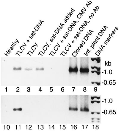
Figure 6.
Encapsidation of TLCV sat-DNA by helper virus coat protein. TLCV and TLCV sat-DNA associated with encapsidated viral particles was detected by immunocapture PCR. PCR was carried out with TLCV-specific (lanes 1–8) or TLCV sat-DNA-specific primers (lanes 10–17). The negative photographic image of the ethidium-stained PCR products are shown. Lanes 1–6 and 10–15 show PCR products obtained with template immunocaptured from extracts of Datura plants agroinoculated with TLCV or TLCV+TLCV sat-DNA, respectively. Extract shown in lanes 4 and 13 was spiked prior to immunocapture with total nucleic acid containing TLCV sat-DNA extracted from twice the weight of tissue used for immunocapture. Negative controls consisted of extract immunocaptured with an unrelated antibody against cucumber mosaic virus coat protein (lanes 5 and 14) or with no antibody (lanes 6 and 15). Control PCR reactions were carried out with using either cloned DNA templates (lanes 7 and 16) or DNA isolated from TLCV-infected plants as described in Fig. 1 (lanes 8 and 17).

