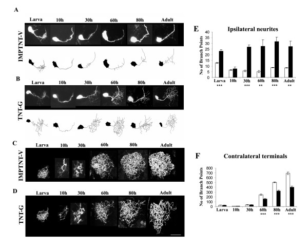
Figure 6.
Effect of expression of TeTxLC on the remodeling of the CSD neuron. The developmental profile of the CSDn in animals where the (a, c) inactivated (UAS-IMPTNT-V/+; RN2-Flp, Tub-FRT-CD2-FRT-Gal4, UAS-CD8GFP/+) and (b, d) activated (UAS-TNT-G/+; RN2-Flp, Tub-FRT-CD2-FRT-Gal4, UAS-CD8GFP/+) forms of TeTxLC have been expressed. Animals where a unilateral flipout had occurred were analyzed in detail at the larva, 10 hour APF, 30 hour APF, 60 hour APF, 80 hour APF and adult stages. Scale bar for all images = 30 μm. The dendritic trees from preparations where inactivated (a) and activated (b) TeTxLC have been ectopically expressed are schematized. The presynaptic terminals in the contralateral lobe where IMPTNT-V and TNT-G were expressed are shown in (c) and (d), respectively. The branch points were quantified as described in Additional file 3 and represented in the histograms in (e) for the ipsilateral lobe and (f) for the contralateral lobe. Five preparations were analyzed at each data point; white bars represent UAS-IMPTNT-V/+; RN2-Flp, Tub-FRT-CD2-FRT-Gal4, UAS-CD8GFP/+; black bars represent UAS-TNTG/+; RN2-Flp, TubFRT-CD2-FRTGal4, UAS-CD8GFP/+. P-values were calculated using the unpaired Student's t-test; **P < 0.001, ***P < 0.0001.

