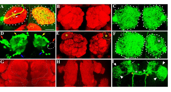
Figure 7.
Effect of ablation of subsets of neurons within the olfactory circuit on the development of the CSD neuron. (a) Antennal lobes (demarcated with dotted lines) of lz3; +/+; Antp/RN2-Flp, Tub-FRT-CD2-FRT-Gal4, UAS-CD8GFP stained with anti-GFP (green) and mAbnc82 (red). lz3; +/+; Antp/+ animals lack ORNs projecting from the third antennal segment. As in the wild-type (Figure 1a), the CSDn in this genetic background sends a few dendrites into the ipsilateral lobe (small arrows) and terminates by extensive branching in the lobe contralateral to the cell body. The glomeruli are less well demarcated by mAbnc82 staining compared to control brains shown in (b). (c) RN2-Flp, Tub-FRT-CD2-FRT-Gal4, UAS-CD8GFP where terminals of both CSDn are labeled within the antennal lobes. (d-f, h, i) Brains of animals that had been fed with HU, stained with anti-GFP (green) and mAbnc82 (red). (d, e) GH146-Gal4, UAS-GFP. (d) Cell bodies of the PNs lie in three clusters located anterodorsal (blue arrows), posterior (yellow arrows) and lateral (red arrows) to the antennal lobe. A subset of the lateral cluster neurons (dotted region) is absent in one lobe. (e) This results in the absence of a glomerulus denoted with (*-DA1) on the affected side (yellow dotted lines). (f) RN2-Flp, Tub-FRT-CD2-FRT-Gal4, UAS-mCD8GFP animals treated as the GH146-Gal4, UAS-GFP animals described above. Animals with one lobe reduced in size were selected and the terminal arbors from bilateral flip-outs examined. The affected side shows significantly less branching, particularly over the region where the DA1 is positioned (asterisk; compare with (c)). (g) Mushroom bodies are indicated by dotted lines in control (no HU) preparations. (h, i) In HU treated animals where the mushroom bodies were absent (h), the architecture of the CSDn was normal (i). Secondary branches (yellow and white asterisk) that arborize over the calyx of the mushroom bodies (arrows) and the lateral horn (arrowheads) appear normal. Arrows indicate branching at the calyx region and arrowheads the branching at the lateral horn. Scale bar = 20 μm.

