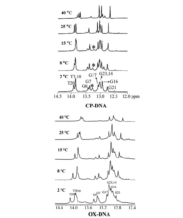
FIGURE 5.

Expanded imino region from 1D 1H NMR spectra of the CP–DNA (top) and OX–DNA (bottom) (26) duplexes recorded in H2O buffer at various temperatures. The positions of the nucleotides in the 12-mer duplexes that give rise to the resonances are indicated. The asterisk in the 1D 1H NMR spectra of the CP–DNA adduct indicates the terminal guanine (G24). The experimental temperatures are shown at the left.
