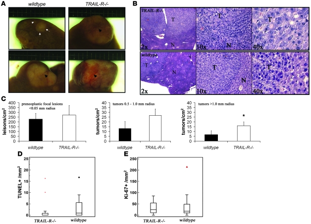
Figure 8. Loss of TRAIL-R renders mice susceptible to DEN-induced hepatocarcinogenesis.
(A and B) TRAIL-R–/– mice (n = 10) injected with DEN (0.30 mmol/kg body weight) at 7 days of age showed an increased liver tumor load compared with WT littermates (n = 10) at 10 months after treatment. (A) Gross observations of small (top row, demarcated with arrows and arrowheads) and larger liver tumors in DEN-treated animals (bottom row, demarcated with arrowheads). (B) Microscopy of H&E-stained histological sections shows the location of a liver tumor (T) with surrounding normal (N) liver tissue. (C) Detailed histological analysis of livers (>0.5 cm2 liver area analyzed, excluding macroscopic lesions) from randomly selected littermates (n = 3/genotype) shows an increased number of lesions exceeding a radius of 1.0 mm in TRAIL-R–/– animals. *P < 0.05, Student’s t test. Means ± SD are shown. (D and E) Immunohistochemical analysis of proliferation (Ki-67 labeling; E) and cell death (TUNEL staining; D) in HCCs from WT (n = 17) and TRAIL-R–/– (n = 17) animals treated with DEN show reduced TUNEL labeling in TRAIL-R–/– HCCs (Mann-Whitney U test, P < 0.05) compared with WT HCCs, whereas similar levels of Ki-67–positive cells were seen in HCCs of both genotypes.

