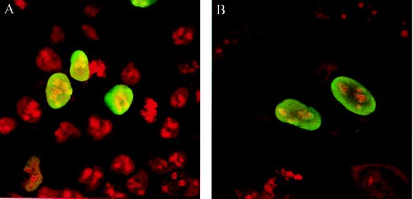
Figure 2.
Nuclear localization of the VirD2 protein in human cells. (A) HEK 293 cells. (B) HeLa cells. The cells were transfected transiently (HEK 293) or were microinjected (HeLa) with pD2. VirD2 protein was visualized by indirect immunofluorescence using anti-VirD2 antibody and fluorescein isothiocyanate-conjugated anti-rabbit IgG antibody. The cells were fixed and stained with propidium iodide.

