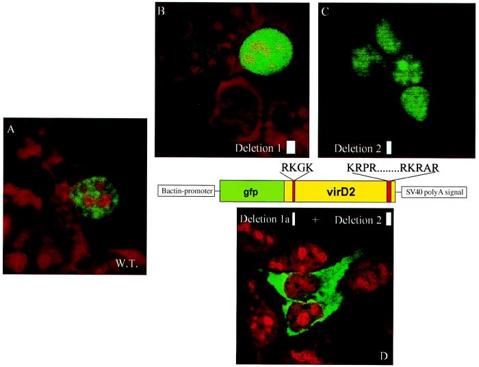
Figure 3.
Subcellular localization of the VirD2 NLS mutant proteins fused to GFP, in HEK 293 cells. (A) Wild-type VirD2 protein fused to GFP (cells were transfected transiently with pGFP-D2). (B) N-terminal NLS mutant VirD2 protein (cells were transfected transiently with pGFP-PstD2. (C) C-terminal NLS mutant VirD2 protein (cells were transfected transiently with pGFP-S132). (D) Mutant VirD2 protein lacking both N- and C-terminal NLS (cells were transfected transiently with pGFP-S155). Cells were fixed and stained with propidium iodide.

