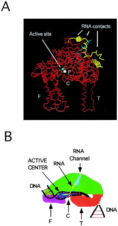
Figure 7.
(A) α-Carbon backbone (red) representation of the crystal structure of T7 RNAP (10). The coordinates in Brookhaven PDB file (4RNP) were displayed using an INSIGHTII program and a Silicon Graphics workstation. The major cross-link site is in cyan and the minor site is in yellow. The white and blue spheres are D537 and D812, respectively, in the active site. (B) A model of transcription complex based on the shape of T7 RNAP. C, cleft; P, palm is in blue; T, thumb is in orange; F, fingers are in violet; the DNA is in black while the base pairs are in red; RNA is in yellow. The proposed RNA channel is shown in light blue.

