Figure 1.
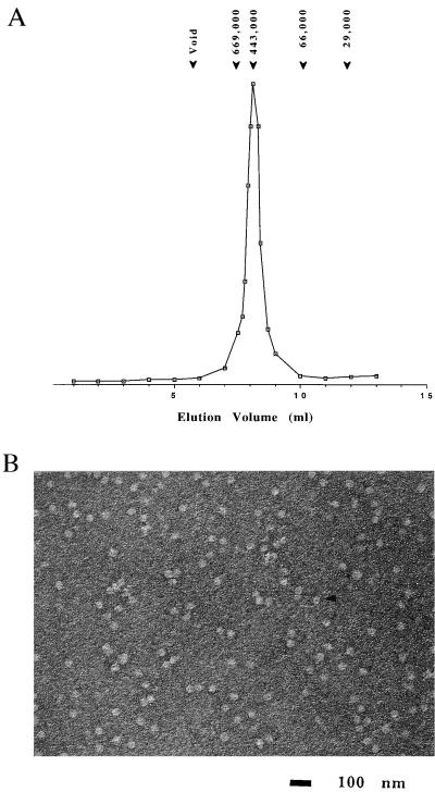
SEC and electron microscopy (EM) of Mj HSP16.5. (A) SEC was performed by using a Tosohaas TSK G4000SW column as described in Materials and Methods. Purified Mj HSP16.5 elutes as a single peak of molecular mass ≈440 kDa. The column was standardized with the following markers as indicated above the figure: carbonic anhydrase (29 kDa); albumin (66 kDa); apoferritin (443 kDa); thyroglobulin (699 kDa). (B) Negative stain EM image of Mj HSP16.5 (1 μM) appears as small particles ≈15–20 nm in diameter. The arrow shows a particle displaying a hole. (Bar = 100 nm.)

