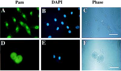
Figure 5.
Subcellular localization of Pam. Immunofluorescence microscopy was performed on normal human aortic endothelial cells with antibody directed against Pam. (A) and (D) Immunofluorescence with Pam-specific antiserum. (B) and (E) Staining with 4′,6-diamidino-2-phenylindole (DAPI). (C) and (F) Phase contrast microscopy. The bars represent 40 μm in C and 20 μm in F.

