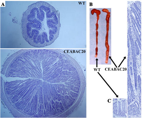
Figure 6. Massive colonic tumors in 3 month-old CEABAC20 mice.
A) Cross sections of colon; note the much larger size of the CEABAC20 vs WT colons. B) Dramatic increase of colon mass and absence of fecal pellets in CEABAC20 over the entire colon. C) Colonic crypts; note the dramatic lengthening of intensely stained crypts in CEABAC20. Magnification: 40× (A), 100× (C). Staining: hematoxylin (A, C).

