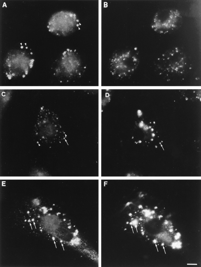
Figure 8.
Simultaneous localization of ankyrin-3 and endocytically incorporated, FITC-labeled dextran. (A, C, and E) Ank3-R1 antibody. (B, D, and F) FITC-dextran. Cells were pulsed for 15 min with 1 mg/ml FITC-dextran, and then washed thoroughly and chased in BMM media for 0 h (A and B), 6 h (C and D), and 24 h (E and F). Arrows and arrowheads indicate ankyrin-3–positive vesicles that do or do not colocalize with FITC-dextran, respectively. Note that at 6 h a small subset of ankyrin-3–positive vesicles contain FITC-dextran, but complete coincidence between ankyrin-3 and vesicular FITC-dextran does not occur until the 24-h chase period. Bar, 10 μm.

