
Figure 3.
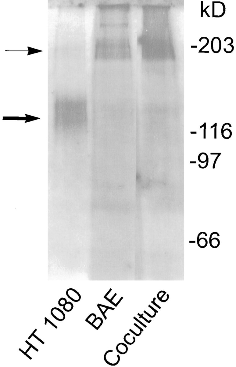
Presence of LTBP-1 in matrix digests prepared from HT 1080, BAE, and cocultures of BAE and BSM cells. Subconfluent cultures of HT 1080 and BAE cells and cocultures of BAE and BSM cells were pulsed with [35S]cysteine for 3 h and chased overnight. Matrices were prepared, followed by digestion with 0.3 U/ml of plasmin. Digests were immunoprecipitated using Ab 39 serum followed by protein A–agarose. Immunoprecipitates were analyzed by SDSPAGE followed by fluorography. ➞ , 120–140-kD LTBP-1 matrix fragments. → , 180– 210-kD LTBP-1 matrix fragments.