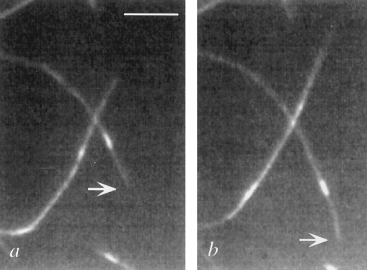
Figure 2.
Movement of polarity-marked microtubules in the plus direction. The microtubule minus end is nearest the rhodamine seed and is indicated by the arrow. (a) Microtubule position at t = 0. (b) Microtubule position at t = 6 min. The velocities are not as fast as those in the non–fluorescence-based assays, presumably owing to photodamage. Bar, 5 μm.

