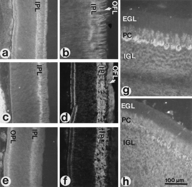
Figure 7.
Localization of CALEB in sections of the developing retina and cerebellum. Cryostat sections were stained indirectly by mAb 4/1 directed to CALEB (a, b, d, and g), by mAb M1 to TN-C (c, f, and h) or mAb 23-13 to TN-R (e). Staining was visualized by fluorescence. Retinas were from 8-d-old (a, b, c, and e) or from 12-d-old chicken embryos (d and f). Cerebella were from 18-d-old chicken embryos (g and h). b shows the distribution of CALEB in the OFL at a higher magnification as shown in a. EGL, external granular layer; IGL, internal granular layer; PC, Purkinje cells.

