
Figure 6.
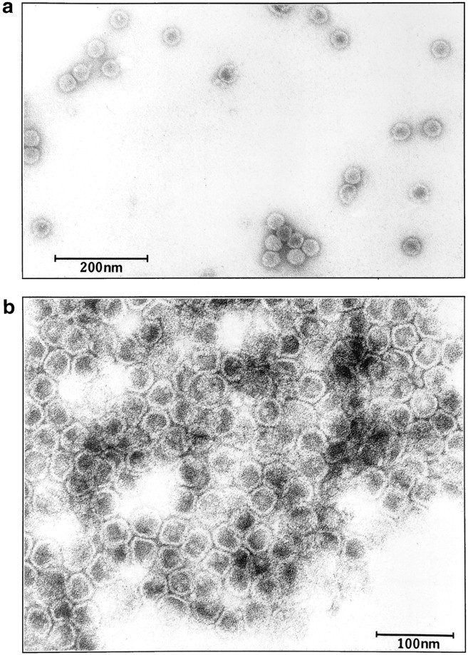
Electron micrograph of purified vesicles. Vesicles were isolated from the second sucrose gradient (fractions 10 and 11). Samples (5 μl) were adsorbed to a carbon-coated grid and examined using a JEOL 1200 EX transmission electron microscope. Bars: (a) 200 nm; (b) 100 nm.