
Figure 1.
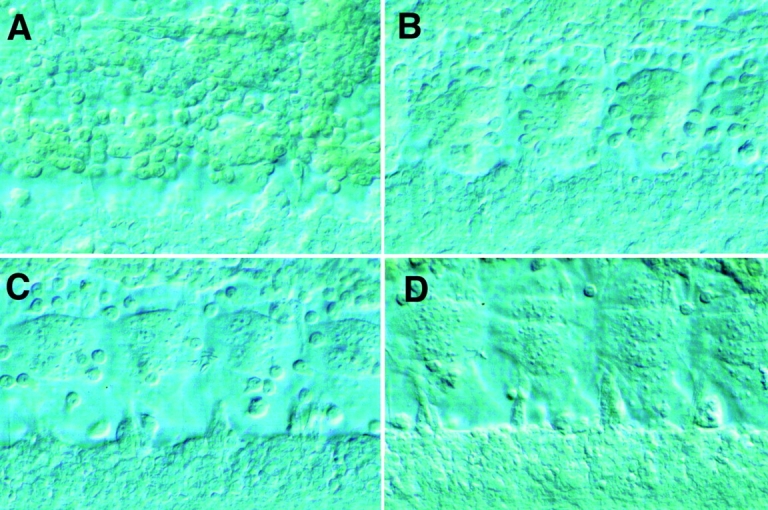
Myoblast fusion in the developing Drosophila embryo. Light level micrographs of myoblast fusion in the ventral muscle region of wild-type Drosophila embryos. Developing muscles are imaged by Nomarski optics, and the plane of focus is close to the epidermis. (A) Wild-type early stage 13 embryo. Small early myotubes are present, with many unfused myoblasts attached to the surface of the myotubes. (B) Wild-type stage 14 embryo. (C) Wild-type stage 15 embryo. Myotubes are substantially larger, with few unfused myoblasts remaining. (D) Wild-type stage 16 embryo.