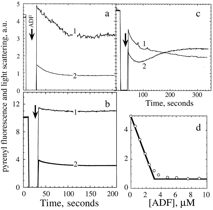
Figure 3.
ADF1 binds to labeled F-actin with concomitant quenching of fluorescence followed by partial depolymerization. (a) Simultaneous recordings of light scattering (1) and pyrenyl fluorescence (2) upon addition of 3 μM ADF (arrow) to a 4.5 μM 100% pyrenyl-labeled F-actin solution. The curves are normalized by adjusting the light scattering and fluorescence intensities of F-actin recorded before addition of ADF to the same maximum level and subtracting the intensities corresponding to G-actin. (b) Same as in a, but 7.5 μM ADF was added to 13.5 μM fully labeled pyrenyl–F-actin. (c) Same as in a, with a 0.5% labeled pyrenyl– F-actin solution. (d) The extent of quenching of fluorescence of 100% labeled pyrenyl–F-actin (3.6 μM) upon binding ADF is plotted versus ADF concentration.

