
Figure 9.
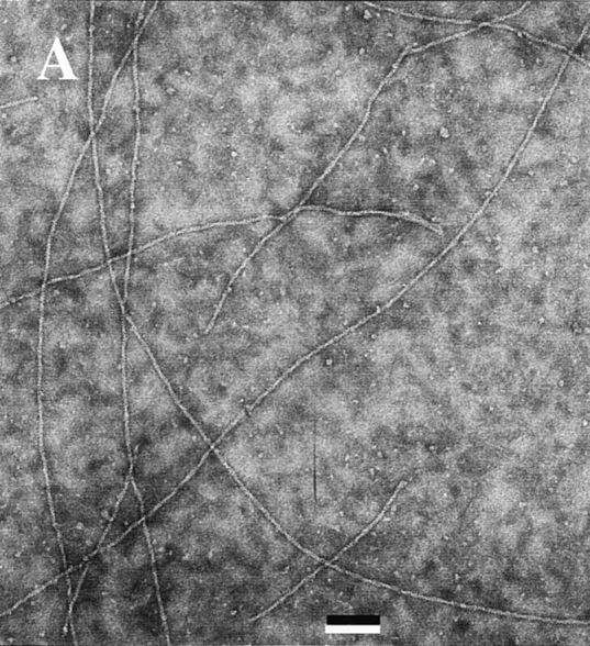
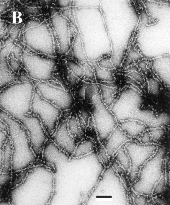
Electron micrographs of F-actin and ADF–F-actin filaments. A solution of 4 μM F-actin polymerized in the absence (A) and in the presence (B) of 5 μM ADF1 in physiological ionic strength buffer, pH 7.8, was deposited on the grid using a largely truncated pipet tip and processed for negative staining as described (Carlier et al., 1994). Bar, 0.1 μm.