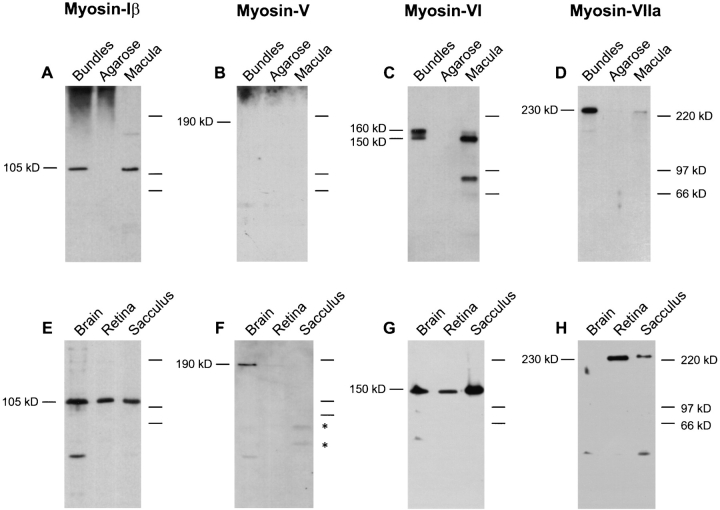
Figure 1.
Protein immunoblot detection of unconventional myosin isozymes expressed in frog hair bundles and tissues. (Top panels) Frog saccular hair bundles were isolated by the twist-off method (Gillespie and Hudspeth, 1991). Bundles, ∼40,000 hair bundles (21 saccular equivalents). Agarose, ∼2 mg of agarose, from agarose adjacent to purified bundles but free of tissue, as a control. Macula, sensory epithelia cells (without peripheral cells, basement membrane, or nerve) remaining after bundle isolation. Protein for ∼1.0 sensory epithelium (2,000 hair cells and 4,000 supporting cells) was loaded. Proteins were separated by SDS-PAGE, transferred to PVDF membranes, and probed with antibodies specific for myosin-Iβ (A and E), -V (B and F), -VI (C and G), and -VIIa (D and H), as described in the text. (Bottom panels) Total protein (10 μg) from brain, retina, and whole saccule was loaded. On low cross-linker gels such as these, myosin-Iβ migrates with an estimated molecular mass of ∼105 kD. Asterisks in F indicate saccular proteins that cross-react with the 32A antibody. Detection was with the following antibodies: (A and E) rafMIβ; (B and F) 32A; (C and G) rapMVI; (D and H) rahMVIIa.

