
Figure 4.
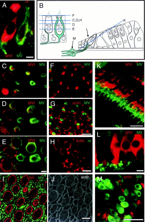
Localization of myosin-V in guinea pig utricle and cochlea and in frog saccule. All images are single optical sections. (A) Double labeling showing myosin-V (green) in neuronal processes contacting type II (left) and type I (right) hair cells in guinea pig utricule; hair cells also labeled for myosin-VI (red) to visualize cell bodies. (B) Depiction of guinea pig utricule on left and cochlea on right. Optical section levels for some images are indicated. (C–E) Confocal z-series through utricular hair cells labeled for myosin-V (green) and myosin-VI (red), showing rings of myosin-V labeling associated with calyces enveloping type I hair cells. (F and G) Labeling of utricular cells for myosin-V (green) and actin (red), showing myosin-V staining is absent from stereocilia in F and from circumferential actin belts in G. (H) Nonimmune control labeling (green) of utricular hair cells at the level of the circumferential actin belt; actin labeling is shown in red. (I and J) Apical surface of bullfrog saccule, triple labeled for myosin-V (green, I), myosin-VI (red, I), and actin (J); myosin-V labeling is associated with supporting cell apical surfaces. (K) Section through guinea pig cochlea, stained for myosin-V (green) and -VI (red). Myosin-V labeling is associated with neuronal processes contacting the bases inner hair cells. (L) High magnification view of bouton endings contacting cochlear inner hair cells; stained for myosin-V (green) and -VI (red). (M) Spiral ganglion neurons stained for myosin-V (green) and actin (red). Bars: (A, C–J, and L) 10 μm; (K and M) 50 μm.