
Figure 6.
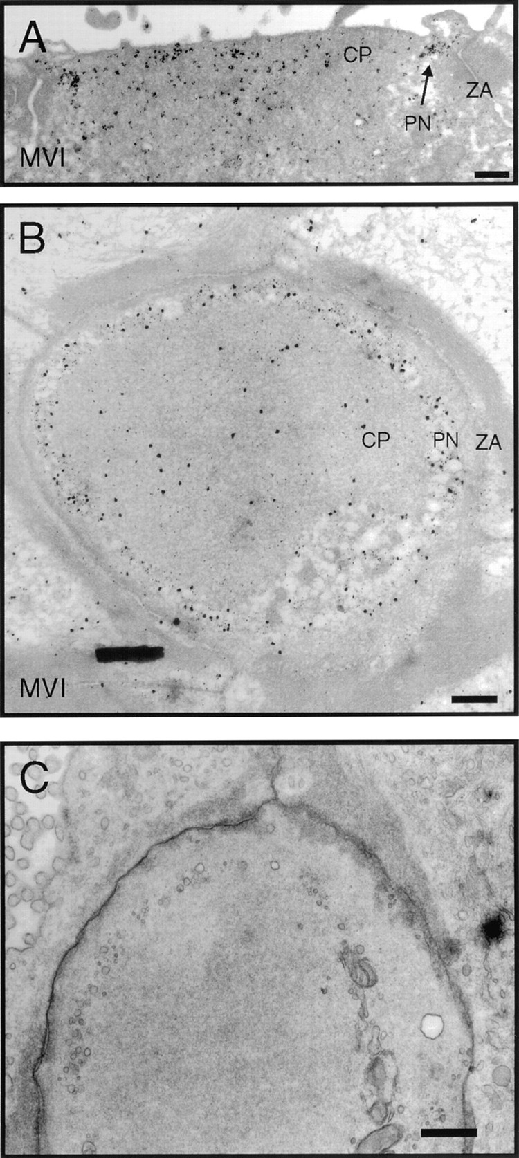
Immunoelectron microscopic localization of myosin-VI in frog saccule. (A) Vertical cross-section through the cuticular plate region showing pericuticular necklace labeling (PN) between cuticular plate (CP) and circumferential actin belt at the zonula adherens (ZA). (B) Horizontal section through the cuticular plate and zonula adherens. Label in the hair cell at this level is strongest in the regions not occupied by actin. (C) Same level as B but with more rapid fixation and without antibody labeling with its extensive tissue extraction. Cytoplasmic vesicles are visible in the pericuticular necklace region. Bars: (A–C) 1 μm.