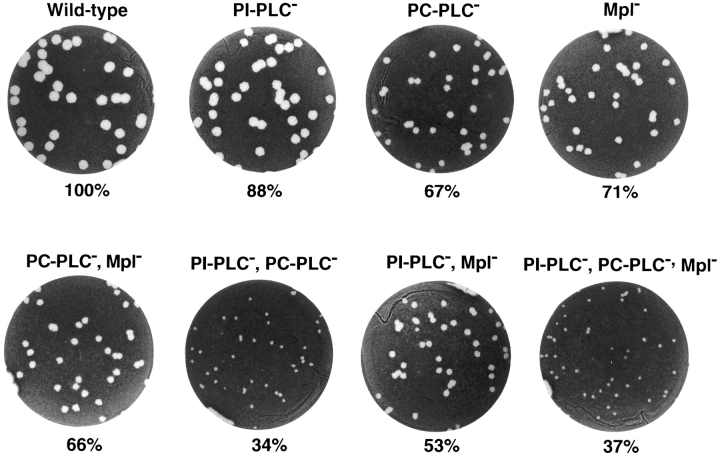
Figure 2.
Formation of plaques in infected L2 mouse fibroblasts. L2 cells were infected with strain 10403S and isogenic mutants. Cell monolayers were stained with neutral red at 4 d after infection, highlighting clear areas of dead cells resulting from L. monocytogenes intracellular growth and cell-to-cell spread. The mean plaque diameters were calculated from several individual experiments (see Table II) and are indicated below each well.

