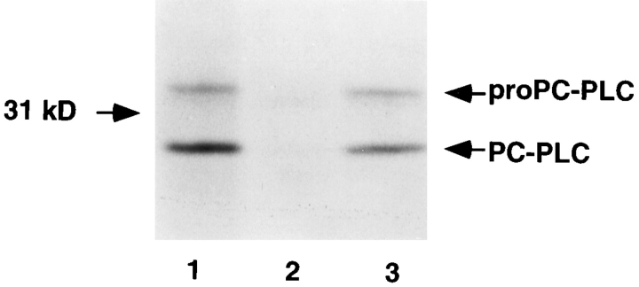
Figure 3.
Detection of PC-PLC from lysates of infected cells. J774 cells were infected with strain 10403S and isogenic mutants. At 4 h after infection, cells were pulse labeled for 30 min with [35S]methionine. Immediately after the pulse, the cells were lysed, PC-PLC was immunoprecipitated using affinity-purified antibodies, and proteins were fractionated by SDS-PAGE. PC-PLC was detected by fluorography. (Lane 1) 10403S; (lane 2) DP-L1935 (PC-PLC−); (lane 3) DP-L2296 (Mpl−). Number of colony forming units (CFU) per sample: 2.8 × 10⁷ (lanes 1 and 3), and 2.6 × 10⁷ (lane 2).

