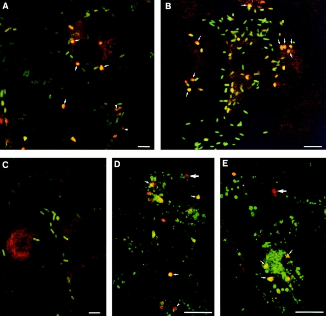
Figure 5.
Immunolocalization of PC-PLC in infected cells. J774 cells were infected with strain 10403S (A, B, and D), or isogenic mutants DP-L1935 (PC-PLC−) (C) and DP-L2296 (Mpl−) (E). Cells were fixed in formalin at either 5 (A and C–E) or 6 h after infection (B), stained for immunofluorescence analysis as described in Materials and Methods, and analyzed by confocal microscopy (0.5-μm sections). (A–E) PC-PLC (rhodamine); (A–C) L. monocytogenes (fluorescein); (D and E) Lamp1 (fluorescein). Overlapping of both fluors (rhodamine and fluorescein) generates yellow. (A, arrows) Examples of bacteria/PC-PLC colocalized staining suggesting vacuolar localization; (arrowheads) PC-PLC staining at the septum of dividing bacteria. (B) In this experiment, cells were infected with 10-fold less bacteria to facilitate identification of primary (highly infected central cell) and secondary infected cells (peripheral cells). The background of this particular field was increased by computer to facilitate visualization of the cells. (Arrows) Examples of bacteria/PC-PLC colocalizing in three different secondary infected cells. (D and E) Large arrows show examples of PC-PLC–containing vacuoles with no Lamp1 staining, whereas small arrows show examples suggesting advanced fusion events between PC-PLC–containing vacuoles and Lamp1-positive endosomes. Arrowhead at the bottom of D shows a PC-PLC–containing vacuole surrounded by Lamp1. Bars, 10 μm.

