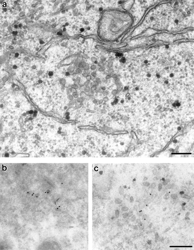
Figure 9.

Morphology of mitotic Golgi clusters by electron microscopy. NAGFPHeLa cells were synchronized by sequential blocks with thymidine and aphidicolin to enrich for mitotic cells. After fixation, cells were prepared for (a) Epon or (b and c) immunogold microscopy using antibodies to GFP followed by secondary antibodies coupled to 10-nm gold. The samples in c were postembedded in Epon to better visualize the membranes. Bars, 0.2 μm.
