Figure 3.
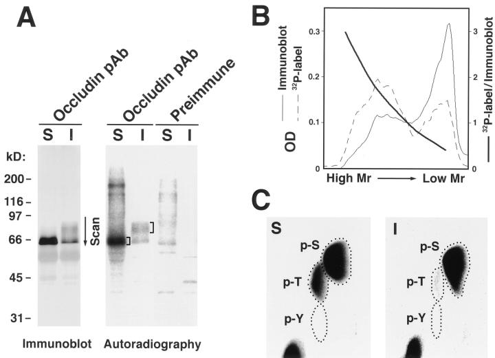
Phosphoamino acid analysis of the NP-40–soluble and -insoluble occludin. (A) Anti-occludin mAb (MOC37) immunoblots (Immunoblot) and accompanying autoradiograms (Autoradiography) of anti-occludin pAb (F5) immunoprecipitates from the NP-40–soluble (S) and NP40–insoluble (I) fractions of confluent MDCK I cells metabolically labeled with [32P]orthophosphate. Control experiments were performed using preimmune serum (Preimmune). (B) Relative specific activity of occludin bands. The region marked by an arrow in the immunoblot and autoradiogram lanes of NP40–insoluble occludin was scanned by densitometry (A, Scan). Relative specific activity of each occludin band was calculated as autoradiogram density/immunoblot density. (C) The marked region in the autoradiogram lane of 32P-labeled NP-40–soluble and -insoluble occludins was excised and processed for phosphoamino acid analysis. The positions of phosphoserine (p-S), phosphothreonine (p-T), and phosphotyrosine (p-Y) were determined by autoradiography through comparison with the ninhydrin staining profiles of unlabeled phosphoamino acid standards. In NP-40–soluble occludin, both serine and threonine residues were phosphorylated (S), whereas in higher M r bands of NP-40–insoluble occludin, serine residues were predominantly phosphorylated with slight phosphorylation of threonine residues (I).

