
Figure 2.

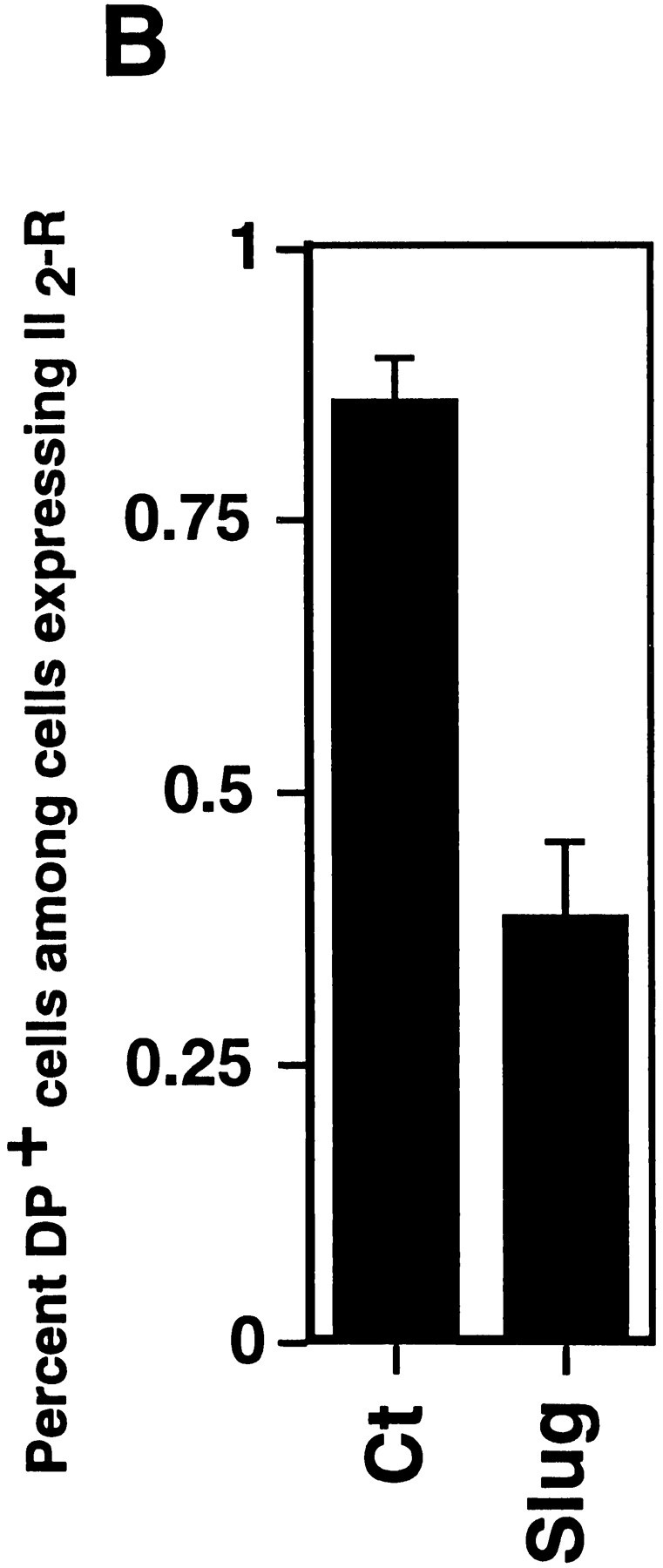
Slug transient transfections. NBT-II cells were cotransfected with vectors containing mouse full-length cDNA for Slug and a truncated (IL-2R) cDNA used as a transfection marker. Alternatively, a control vector pCR 3 (Control) was cotransfected with IL-2R cDNA. After 48 h, cells were fixed and processed for double immunofluorescence using antibodies against IL-2R and antidesmoplakin (A); note that cells expressing the IL-2R transfection marker are positive for desmoplakin in control transfectants and negative in slugtransfected cells. Cells expressing desmoplakin at cell–cell boundaries were counted as desmosome-positive (DP+), i.e., fully epithelial. More than 100 cells expressing IL-2R were analyzed at the same time point for desmosome expression. The number of desmosome-positive cells displaying IL-2R labeling was normalized to the total number of cells expressing IL-2R (B). Bar, 9 μm.