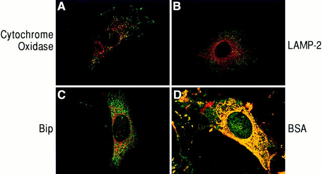
Figure 3.
Confocal micrographs of cells immunostained for PAK1 and cytoplasmic organelle markers. Swiss 3T3 cells in serum were fixed in 100% methanol and costained with anti-PAK1, and antibodies to organelle markers as described in Materials and Methods. (A, B, and D) Red, rhodamine staining of cytochrome oxidase (mitochondrial marker), LAMP-2 (lysosomal marker), and BSA, respectively; green, fluorescein staining of PAK1. (C) Green, fluorescein staining of BiP (endoplasmic reticulum marker); red, rhodamine staining of PAK1; yellow, areas of colocalization.

