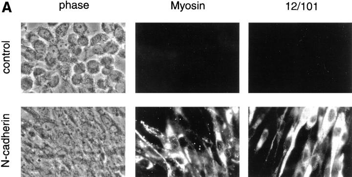
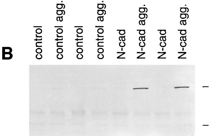
Figure 2.
N-cadherin increases sarcomeric myosin and the 12/101 antigen in aggregated BHK cells. Control cells and cells expressing exogenous N-cadherin were aggregated and examined for the expression of two markers of skeletal muscle differentiation. (A) Cells were aggregated for 24 h, then collected, and placed on slides for 48 h. Sarcomeric myosin was detected with MF20 and the 12/101 antigen was detected with a monoclonal antibody. The apparent differences in the number and morphology between control cells and cells expressing N-cadherin are due to the different cell–cell adhesion properties of the cells (Fig. 3). (B) Two independent clones expressing N-cadherin (N-cad) and two control clones were grown as a monolayer and as aggregates in hanging drops (agg.) for 72 h. The cells were collected and extracted in SDS sample buffer. Extracts were resolved by SDS-PAGE, transferred to nitrocellulose, and immunoblotted with MF20 to sarcomeric myosin. High range molecular weight markers were used and the positions of the 205- and 116.5-kD bands are indicated at the right side of the figure. No increase in myosin or 12/101 was seen in N-cadherin–expressing cells grown in a monolayer.


