Figure 4.
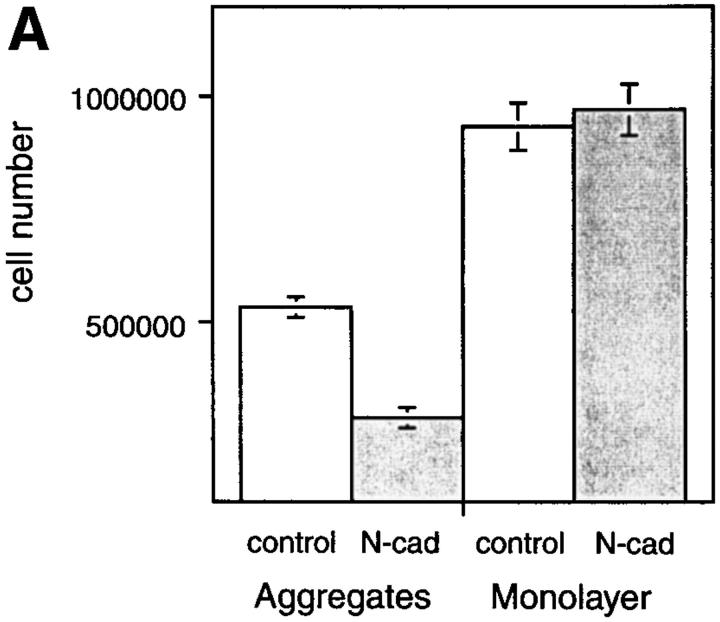
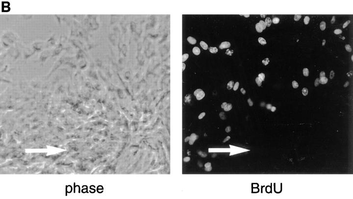
Culturing BHK cells in aggregates inhibits their growth, and this inhibition is enhanced by N-cadherin. (A) An equal number of cells (1.5 × 10⁵) expressing N-cadherin (N-cad) or of control cells (control) was grown for 72 h as hanging drops (aggregates) or as individual drops on tissue culture dishes (monolayer). Cells were harvested and dispersed into single cells with trypsin and counted microscopically using a hemocytometer. Viability was determined by Trypan blue exclusion. Viability was >95% in all of the cultures. The mean of four individual experiments is shown and the standard deviation is indicated. Student's test was used to determine there was no significant difference between the growth of N-cadherin–expressing cells and control cells cultured as monolayers (P = 0.3736). There were significant differences between the growth of control cells in aggregates vs monolayers (P < 0.001), the growth of N-cadherin– expressing cells in aggregates vs monolayers (P < 0.001), and the growth of N-cadherin–expressing cells vs control cells cultured as aggregates (P < 0.001). (B) BHK cells expressing N-cadherin were placed in hanging drops for 24 h to promote aggregation. The cells were collected, placed on slides, and grown for 24 h to permit strong attachment to the slides. The cells were then labeled with BrdU and probed for BrdU using a monoclonal anti-BrdU. The position of the aggregate is indicated by an arrow in each panel.


