
Figure 2.
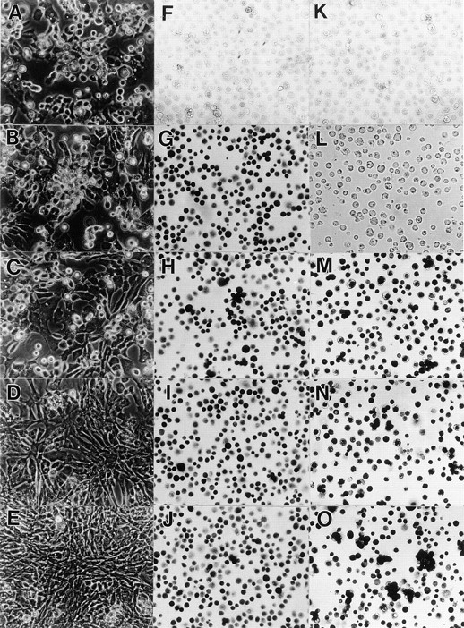
Efficiency of infection in upper sternal chondrocyte cultures by DN-BMPR–encoding viruses. Freshly isolated US chondrocytes were infected with RCAS viruses encoding DN-BMPR-IA (C, H, and M), DN-BMPR-IB (D, I, and N), DN-BMPR-II (E, J, and O), or vector alone (B, G, K, and L) for 3 d in medium 199 containing 10% FBS. The medium was then changed to high glucose DME containing 10% FBS, and cells were cultured for an additional 4 d. On day 7, the cells were harvested and replated in 35-mm dishes at the density of 1.5 × 10⁵ cells/dish, and the microscopic photographs of live cells were taken after 5 d (A–E). Aliquots of the harvested cells were plated onto poly- l-lysine–coated dishes at the density of 2.0 × 10⁶/ml, fixed with 3.7% formaldehyde after 15 min from plating, and stained with antibodies to the RCAS virus gag protein (F–J) or c-myc peptide (L–O) as described in Materials and Methods. A and F represent uninfected cultures. K is a culture incubated with control nonimmune ascites.