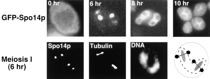
Figure 4.
Relocalization of GFP–Spo14p during meiosis. (Top) Micrographs of living yeast cells harboring GFP–SPO14 2μ (Y969) at the indicated times after induction of meiosis. (Middle) A fixed cell after the meiosis I division showing GFP–Spo14p (Spo14p), antitubulin antibody (Tubulin) and DAPI (DNA) labeling. A schematic representation of the cell is on the right. The circles represent Spo14p, lines represent the spindles, and the ovals represent DNA. The dashed line is the nuclear envelope. (Bottom) A cell expressing GFP–Spo14p (Spo14p) after the meiosis II division stained with DAPI (DNA).


